
Program Report on MSDC’s September Presentation by Tim Rose
by Andy Thompson, MSDC Secretary
This report is provided to encourage readers to visit MSDC’s YouTube channel to view the intriguing video of geologist Tim Rose’s research findings about the airborne minerals and debris of the Canadian wildfires. It lasts 64 minutes including Q & A and you can find it here.
Cindy Schmidtlein, MSDC’s Vice President for Programs, welcomed our club’s sponsor, Tim Rose, who manages the Smithsonian’s Analytical Laboratories of the Department of Mineral Sciences.
The origin of Tim’s presentation, he noted, was his colleague Dr. Ben Andrews, a volcanologist, asking how the Canadian wildfire ash smothering the East coast in June compared with volcanic ash. Tim shared the results of his team’s research which involved collecting samples of the ash and setting up a controlled study of the crystals and debris by using the Smithsonian’s new analytical scanning electron microscope.
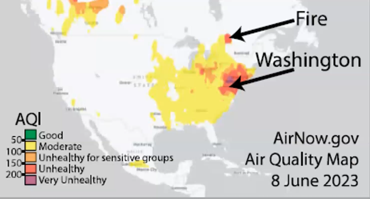
Canadian Wildfire Smoke in the Eastern U.S.
The illustration below shows the extensive range of Canadian smoke that covered the east coast of the U.S. Prior to this event, Code Red on the Air Quality Index had been commonly recognized as the worst-case scenario. But the June smoke registered in DC at an even higher level, as Code Purple.
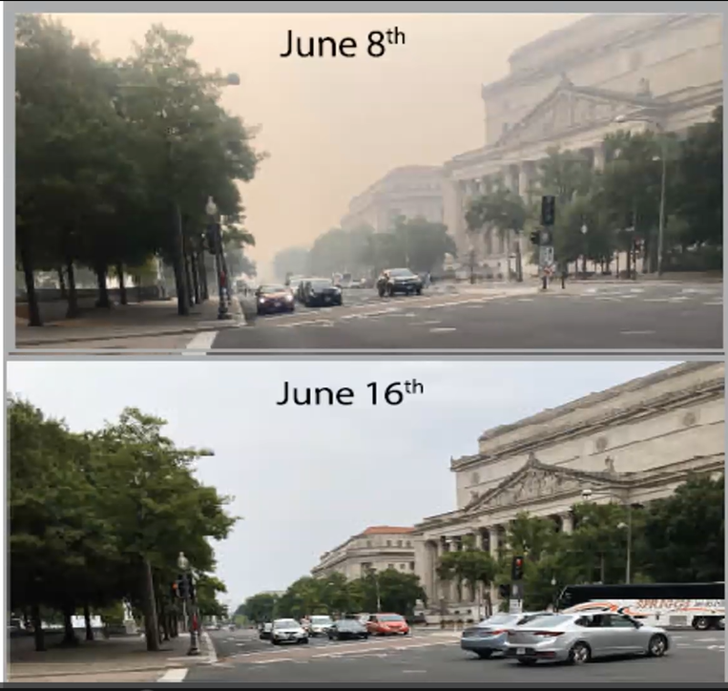
The photos below, taken one week apart on Pennsylvania Ave in Washington, DC, document the problem.
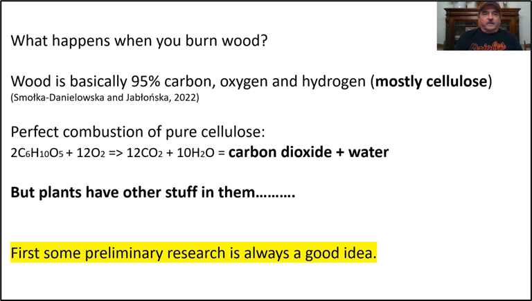
In response to Ben’s question about how this airborne ash compares with volcanic ash, Tim asked: What do we do? His answer: “We do an experiment,” and start by defining the appropriate context and decide on method, including an appropriate, built-in control.
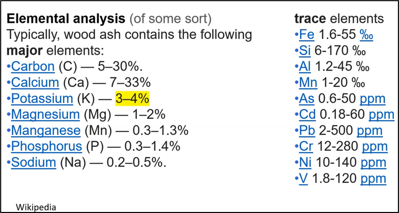
Readily available data provided the answer to the question of what happens when you burn wood. Wood is primarily made up of the three elements, carbon, hydrogen, and oxygen.
The findings from previous basic research identified the following elements in wood ash:
An Important Tool for This Study
The Smithsonian's Department of Mineral Sciences has a scanning electron microscope (SEM) that Tim used to target the ash particles with an electron stream, which resulted in their giving off X-rays. The energy levels of the X-rays revealed what chemical elements were present in the ash grains and the amounts of each.
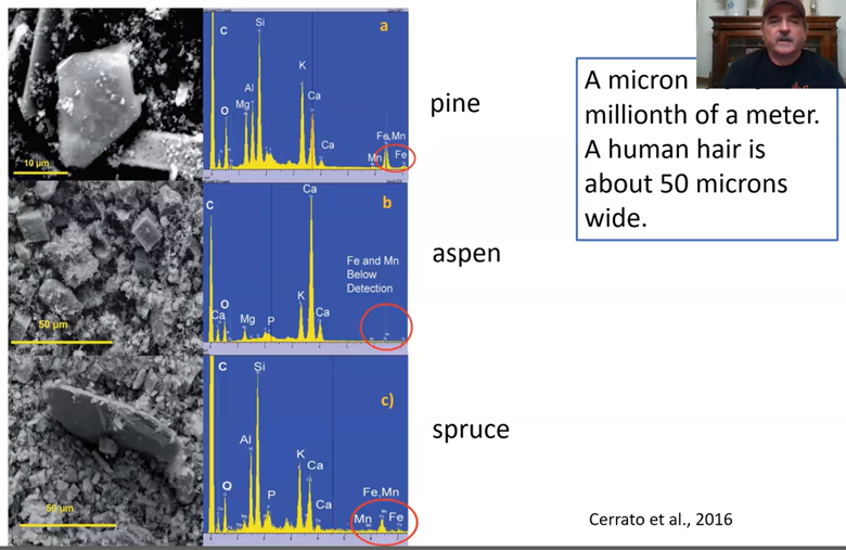
Previously published research, highlighted below, shows images of the very tiny size of the ash grains, crystalline in appearance, smaller than a human hair, found in diverse types of trees. The X-ray spectra identified the quantity of major elements such as carbon, silicon, and calcium, which varied depending on the type of tree from which the ash was derived, as shown below.
Sample Collection and Analysis
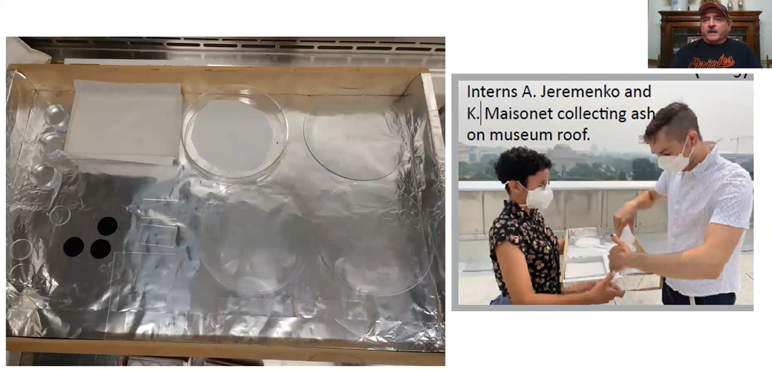
After reviewing the background data described above, Tim and his team were now ready to collect samples of the smoke ash from the roof and east parking lot of the National Museum of Natural History (NMNH). The photo below shows on the far left the 3 black, one-inch diameter “dots” with a sticky surface which acted as a collecting device for obtaining the smoke’s ash crystals. Tim then examined the ash crystals with the Smithsonian’s 18-month-old SEM.
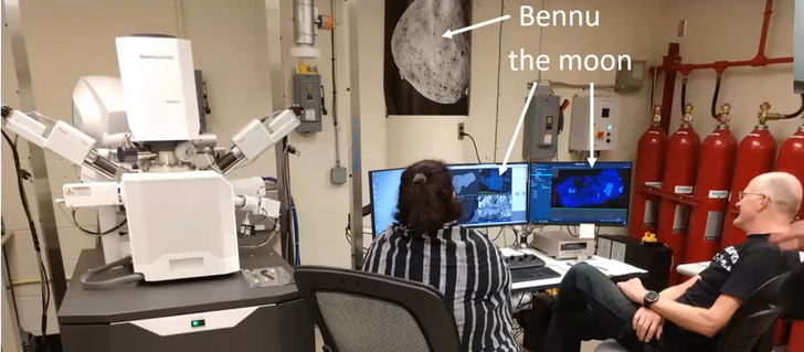
The Smithsonian purchased the SEM scanning machine, shown on the left in the photo below, just 18 months ago, for the specific purpose of analyzing mineral samples anticipated to be collected from the asteroid Bennu. The seven-year Osires-Rex mission to collect samples from the asteroid Bennu culminated with the return to earth of the first-ever Bennu mineral samples on Sunday, September 24th. (Interested readers can see a video of the dramatic touchdown in the Utah desert here.)
Last year, NASA gave samples of lunar material to the Smithsonian allowed the staff to perform analyses using the SEM and to gain familiarity with operation of the SEM.
For comparison, Tim’s curiosity also led him to examine the ash readily available from his personal back-yard smoker. That yielded two types of crystals, one well defined (below) and a second one that was more amorphous.
The scale bar of 10 micrometers (one fifth of the thickness of a human hair), located in the lower right side of the image, indicates that the more-defined crystals were themselves made up of many much smaller cells. Although not important for the purpose of the research, it turned out to be interesting, as explained later in Tim’s presentation, playing an important role for a number of Renaissance masters’ paintings.
Readers who want to get the true flavor of Tim and his team’s research into the crystals and other content of the Canadian smoke can go directly to the video itself by tapping into this link on MSDC’s YouTube channel. It gives access to Tim’s entire presentation, which lasts 64 minutes, including a wonderful question and answer session. But if you want a little more incentive to encourage you to watch the video about this interesting experiment, here are a few more aspects of Tim’s intriguing research and findings.
Results of the Analysis
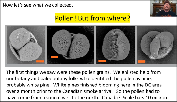
In consultation with his Smithsonian colleagues in paleobotany and botany, Tim determined that the black dots had indeed collected pollen from white pines. That tree genre had stopped blooming in the Washington DC area by April or May.
“This means that we are actually collecting stuff on our little black dots that are actually not from the Washington, DC area. This is pretty good evidence that we actually collected stuff from Canada.”
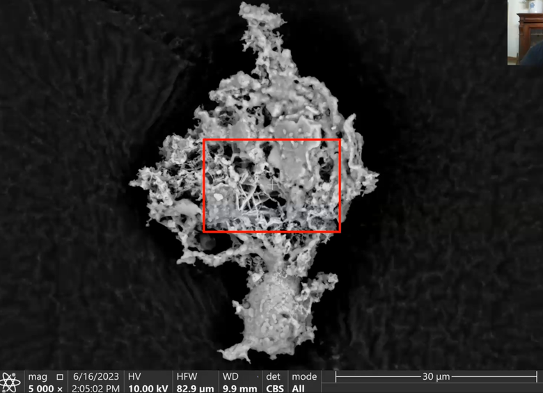
Beyond the pollen, further inspection of the collected material revealed two additional forms from the smoke debris, the first being wispy, amorphous, and even a bit glassy, as shown below.
X-ray analysis showed its elements included the following:
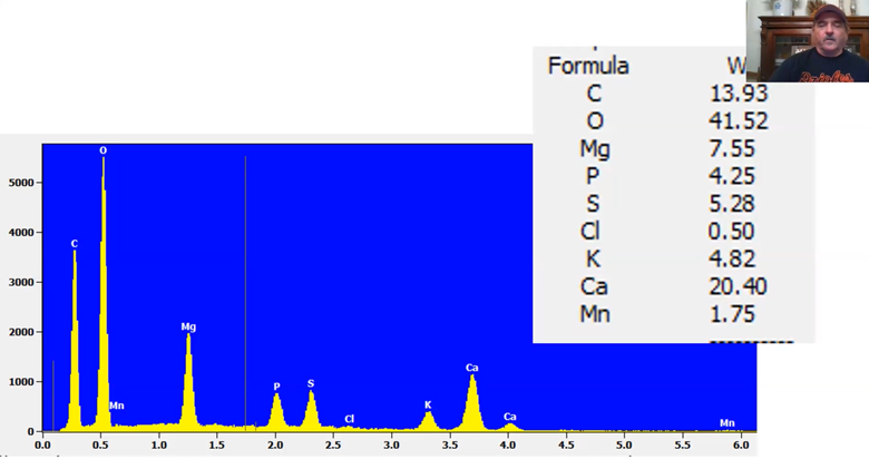
Readers will recall that this image and list of elements is similar in shape and chemical content to what Tim found in the wood ash of his smoker. Both analyses contain the “usual suspects,” oxygen, calcium, carbon, magnesium, etc.
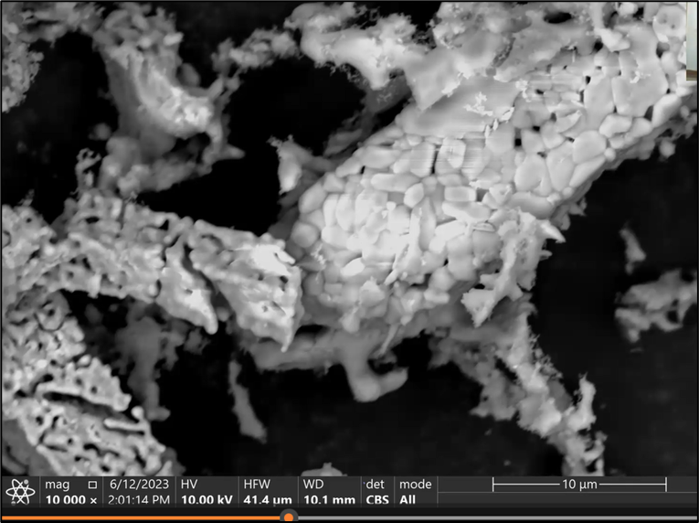
The third form of material found on the black dots, beyond the pollen and wispy material, was the more crystalline and spongy cubes, shown below.
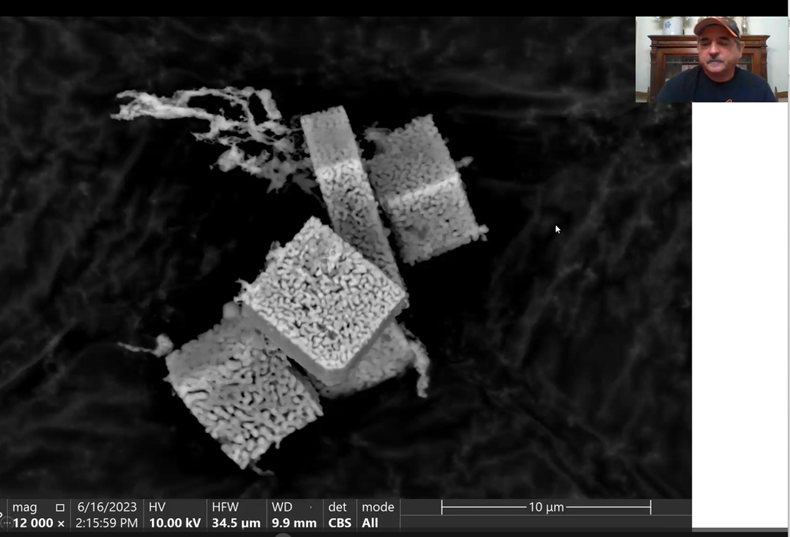
The fact that the above image shows the wispy material along with the crystal cubes suggests they traveled together from Canada and landed together on the black dot collectors. “That means, guys, that we were breathing crystals from Canada.”
Another important question is the role played by the “control,” which is a necessary component of every careful scientific study. Viewers of the video will want to watch for how the control Tim used confirmed that the material collected on the black dots was from Canada and not local to the Washington, DC area.
Yet Another Mystery Revealed
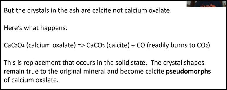
At this point in the study, it seemed that the above pictured cubes were calcium carbonate (CaCO3). But the Smithsonian's Dr. Jeffrey Post, mineralogist and recently retired curator-in-charge of gems and minerals, looked at the images and said they did not look like any calcium carbonate crystals with which he was familiar. So, what are they?
Again, readers are encouraged to view the video tape of Tim’s talk and see what the mysterious cubes were and where they originated. Hint: At the beginning of his talk, Tim talked about “calcium oxalate.”
Tim brought his presentation full circle by revisiting the original question raised by Dr. Ben Andrews: what are the differences between volcanic ash and the Canadian wildfire ash? Tim provided the two photos below of the microscopic crystals found in volcanic ash (on the left) and the Canadian ash on the right.
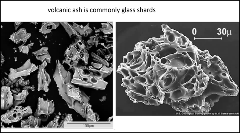
The volcanic ash, Tim explained, is typically a form of glass, with sharp edges, diverse shapes, including with and without bubbles. It has greater density and weight than the Canadian wildfire calcite ash on the right. But both have spiky edges and can cause pulmonary problems if inhaled.
“So that answered (Ben’s) question, but wait, there’s more.”
Tim explained that painting conservators took some sample paint chips from the painting below and used SEM analysis to capture images of their crystal structure.

The following four images are cross-sections of the chips from the above painting.
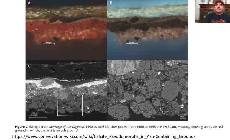
Do you recognize any similarity between the two images at the bottom, C and D, and those Tim discussed above, from the Canadian ash? Hint: The bottom two images were made with the SEM technology and they show spongy looking calcium after calcium oxalate.
After enjoying an extensive and interesting Q and A session, president Kenny Reynolds thanked Tim for his excellent presentation and his Zoom audience showed their appreciation with extensive applause.
The entire program is now available on MSDC's YouTube channel:
Canadian Wildfire Ash: Collection and Study in Washington, DC, June 2023 - Tim Rose - YouTube